Checking Out the Crucial Services Used by a Veterinary Cardiologist: Recognizing Ultrasound and CT Check Techniques
Veterinary cardiologists play an essential role in the wellness of pet dogs by diagnosing and dealing with various heart conditions. They utilize sophisticated imaging strategies, such as heart ultrasound and CT scans, to supply precise evaluations. Each technique has its distinctive advantages and applications. Recognizing these techniques is necessary for pet proprietors seeking the very best treatment for their companions. What variables should pet owners consider when picking in between these diagnostic tools?
The Role of Veterinary Cardiologists in Pet Healthcare
Vet cardiologists play an important function in the health care of pets, focusing especially on identifying and treating heart-related conditions. They have specialized training that allows them to translate complicated analysis tests and recognize different cardiovascular concerns. These professionals utilize advanced methods, such as echocardiography and electrocardiography, to examine heart function and framework accurately.Veterinary cardiologists additionally establish tailored therapy plans that may consist of medications, way of life adjustments, and, in many cases, surgical interventions. Their proficiency reaches informing family pet owners about heart health and wellness, stressing the value of regular exams and early detection of potential problems. Cooperation with basic vets is crucial, as it assures complete care for pet dogs with believed heart concerns. By providing specialized services, veterinary cardiologists greatly enhance the high quality of life for family pets and give comfort for their owners, enhancing the relevance of heart health in general pet wellness.
Usual Cardiac Concerns in Family Pets
Usual heart concerns in pets can greatly affect their health and high quality of life. Heart murmurs, numerous sorts of cardiomyopathy, and genetic heart defects are amongst the most prevalent problems that vets come across. CT Scans For Animals. Understanding these problems is necessary for pet dog proprietors to assure timely medical diagnosis and suitable treatment
Heart Murmurs in Pets
Heart murmurs can be a source of worry for pet owners, they are not always indicative of significant health problems. A heart murmur is an unusual noise generated by unstable blood circulation within the heart. In animals, these whisperings can be triggered by different aspects, including hereditary heart defects, valve concerns, or even anxiety throughout evaluations. Numerous animals with heart whisperings lead typical lives without considerable health and wellness influences. To establish the underlying cause, veterinary cardiologists often employ analysis methods such as echocardiograms and Doppler ultrasounds. Early detection and evaluation are crucial, as they might assist take care of any type of potential cardiac problems efficiently. Animal owners are urged to consult their vet for a complete analysis if a heart whispering is detected.
Cardiomyopathy Kind Explained
Cardiomyopathy encompasses a team of diseases influencing the heart muscle mass, causing jeopardized heart feature in pet dogs. One of the most common kinds consist of expanded cardiomyopathy (DCM), hypertrophic cardiomyopathy (HCM), and limiting cardiomyopathy (RCM) DCM primarily impacts pets, causing the heart to compromise and enlarge, which diminishes its capability to pump blood efficiently. In contrast, HCM is a lot more common in cats, identified by the enlarging of the heart wall surfaces, typically leading to blocked blood flow. RCM, though much less common, happens when the heart muscle comes to be inflexible, limiting its capability to fill up with blood. Each type provides one-of-a-kind challenges in medical diagnosis and treatment, demanding specialized vet cardiological analysis to assure peak management and care for impacted family pets.
Hereditary Heart Flaws
Congenital heart issues represent a considerable group of heart issues in family pets, distinctive from acquired problems such as cardiomyopathy - CT Scans For Dogs. These flaws are structural abnormalities existing at birth, affecting the heart's regular function. Common types consist of license ductus arteriosus, ventricular septal flaws, and pulmonic constriction. Signs and symptoms might vary widely, varying from mild to serious, and can include workout intolerance, coughing, and trouble breathing. Early diagnosis with advanced imaging strategies like ultrasound is crucial for efficient monitoring. Veterinary cardiologists play a vital duty in determining these conditions and recommending ideal treatment choices, which may consist of clinical monitoring or surgical intervention. Identifying hereditary heart problems allows for much better results and boosted lifestyle for influenced pet dogs
Understanding Heart Ultrasound: Just How It Functions
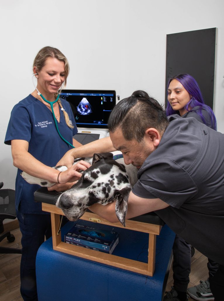
A substantial number of vet methods now use cardiac ultrasound as a necessary analysis device for reviewing heart wellness in animals. This non-invasive method uses high-frequency acoustic waves to develop photos of the heart's structure and feature. During the procedure, a veterinary professional applies a gel to the pet's breast and makes use of a transducer to emit ultrasound waves. These waves jump off the heart and surrounding frameworks, creating real-time images on a monitor.Veterinarians can examine different aspects of cardiac health and wellness, including chamber size, wall motion, and shutoff feature. Additionally, cardiac ultrasound permits the detection of abnormalities such as liquid accumulation and genetic heart issues. This method is important for detecting problems that may not be noticeable through basic radiographs. By supplying comprehensive details about the heart's makeup and efficiency, cardiac ultrasound help in developing efficient treatment plans for animals struggling with heart illness.
The Significance of CT Scans in Detecting Heart Conditions
Just how do CT scans boost the medical diagnosis of heart disease in vet medicine? CT scans provide thorough cross-sectional photos of the heart and bordering structures, allowing veterinarians to envision complex physiological connections. This imaging strategy is particularly helpful in recognizing hereditary heart flaws, cardiac tumors, and problems in capillary. By making use of advanced imaging algorithms, CT scans can analyze heart chamber dimensions and feature, providing a complete view that might be tough to accomplish with traditional methods.Additionally, CT angiography can picture blood circulation and determine areas of stenosis or blockage, which is crucial for planning potential treatments. The rate and accuracy of CT scans also assist in fast medical diagnoses, vital in emergency situations. Ultimately, the consolidation of CT scans right into veterinary cardiology significantly enhances the precision of medical diagnoses, making it possible for targeted therapy plans and improving individual outcomes for pets experiencing heart disease.
Comparing Ultrasound and CT Scan Strategies
While both ultrasound and CT scans are indispensable tools in vet cardiology, they supply unique advantages and constraints that affect their usage in diagnosing heart disease. Ultrasound, or echocardiography, gives real-time imaging of the heart's framework and function, enabling veterinarians to examine heart chambers, valves, and blood circulation. It is especially effective for examining conditions like congestive heart failure and cardiomyopathy. Ultrasound may be restricted in picturing certain anatomical structures due to person dimension or obesity.In comparison, CT scans offer comprehensive cross-sectional photos of the heart and surrounding cells, making them excellent for identifying structural problems, lumps, or vascular concerns. Although CT scans give detailed understandings, they require sedation and might include radiation direct exposure. Eventually, the selection in between ultrasound and CT scans depends on the details medical circumstance, the individual's problem, and the info required for an exact medical diagnosis.
Treatment Choices Offered Through Vet Cardiology
Veterinary cardiology provides a variety of treatment choices tailored to resolve numerous heart disease in animals. Treatment strategies commonly start with way of life alterations, consisting of diet modifications and exercise adjustments, targeted at improving total heart health and wellness. Medications play a crucial function, with cardiologists prescribing medications such as diuretics, beta-blockers, and ACE preventions to handle signs and boost cardiac function.In more extreme situations, interventional treatments, such as balloon valvuloplasty or stent positioning, might be needed to relieve blockages or enhance blood circulation. For particular hereditary heart issues, medical options may be discovered to fix structural problems. Additionally, recurring tracking and follow-up treatment are necessary components of a detailed treatment plan, enabling timely changes based on the family pet's response to therapy. Generally, veterinary cardiology concentrates on providing effective, personalized like optimize the health and wellness and wellness of animal individuals with heart disease.
How to Prepare Your Pet for a Cardiac Examination
Preparing a pet for a heart assessment is vital to assure exact results and a smooth procedure. Proprietors need to initially set up the consultation with the vet cardiologist and review any kind of particular needs or issues. It is a good idea to keep food for at the very least 12 hours prior to the assessment, as this assists improve imaging high quality during treatments like ultrasound or CT scans.Additionally, keeping a tranquil environment on the day of the visit can help in reducing the family pet's anxiety. It is useful to bring along any type of pertinent medical records, including previous examinations and medications (Board Certified Veterinary Cardiologist). Owners must likewise ensure that their animal is comfy and leashed during transportation to the center. Familiarizing themselves with the evaluation procedure can relieve fears and help in asking notified inquiries throughout the assessment. By complying with these steps, proprietors can contribute greatly to the performance of the cardiac evaluation
Regularly Asked Inquiries
The length of time Does a Heart Ultrasound or CT Check Take?
The period of a heart ultrasound generally varies from 30 to 60 minutes, while a CT scan may take about 15 to 30 minutes. Factors such as the person's condition can influence these time estimates.

Exist Any Kind Of Threats Linked With These Analysis Treatments?

Can I Remain With My Family Pet During the Procedure?
The veterinary center's policy commonly determines whether pet owners can continue to be throughout procedures. While some clinics urge owner presence for convenience, others might require splitting up to assure security and perfect conditions for diagnostic imaging.
Just how much Do These Analysis Tests Typically Price?
The expenses of analysis examinations, such as ultrasound and CT scans, normally vary based on area and facility. Normally, rates range from a few hundred to over a thousand dollars, mirroring the intricacy and technology involved.
What Is the Recuperation Process After a Cardiac Evaluation?
The recovery process after a heart assessment includes keeping track of the pet dog for any immediate responses, guaranteeing comfort, and restricting exercise. Veterinarians usually supply post-evaluation guidelines to assist pet owners during this necessary recovery period. Heart murmurs, numerous types of cardiomyopathy, and congenital heart issues are among the most widespread problems that veterinarians encounter. A heart whispering is an uncommon audio generated by stormy blood circulation within the heart. Cardiomyopathy incorporates a group of illness impacting the heart muscle mass, leading to compromised cardiac feature in pets. Congenital heart flaws stand for a considerable classification of heart problems in pet dogs, distinct from obtained conditions such Cancer Veterinary Near Me as cardiomyopathy. Ultrasound, or echocardiography, provides real-time imaging of the heart's structure and feature, allowing veterinarians to assess heart chambers, shutoffs, and blood flow.